Medical Imaging Machine Learning: A Guide to AI-Powered Diagnostics
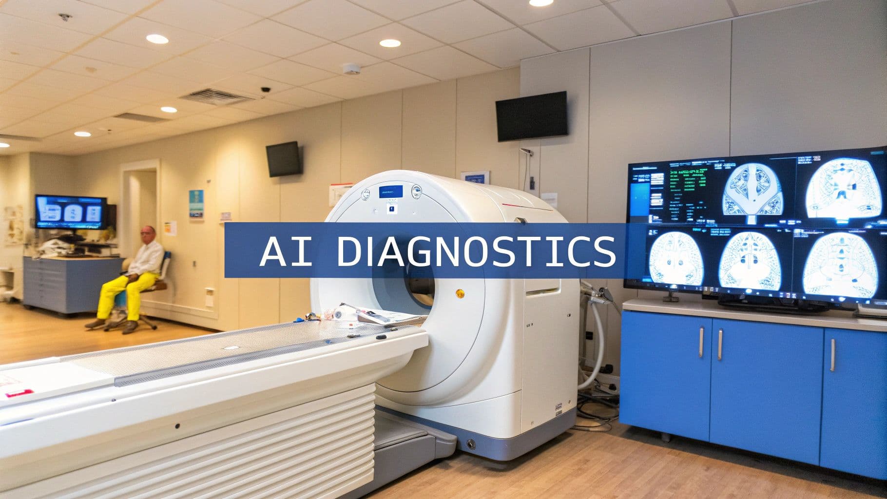
Think of medical imaging machine learning as a highly trained digital apprentice for clinicians. It’s designed to analyze scans like MRIs, CT scans, and X-rays with superhuman precision, catching subtle patterns the human eye might overlook. The goal isn't replacement but collaboration—accelerating diagnoses, sharpening accuracy, and ultimately improving patient outcomes.
A New Era of Diagnostic Insight
Imagine giving a specialist a second set of eyes—eyes that never get tired, never get distracted, and have reviewed millions of scans. That’s the essence of medical imaging machine learning.
These sophisticated algorithms are trained on vast, anonymized libraries of medical images. By processing countless examples of everything from healthy tissue to the earliest signs of disease, they learn to recognize the visual signatures of different conditions. They can spot a tiny lung nodule or a hairline fracture that might otherwise go unnoticed. This isn't about taking over; it's about augmenting the incredible expertise of clinicians.
The sheer volume of imaging data produced today is staggering, creating a significant bottleneck in many healthcare systems. By flagging suspicious findings and handling routine analysis, these tools help radiologists and other specialists focus their energy on the most complex and critical cases. It’s a powerful way to manage the data deluge and deliver faster, more precise care.
Why This Matters Now
This shift couldn't come at a better time. As imaging technologies get more powerful, each scan generates more data than ever before, making manual review a monumental task. Machine learning steps in to process this complexity efficiently, turning raw pixels into actionable clinical insights.
The momentum is undeniable. The global market for AI in medical imaging was valued at around USD 1.36 billion and is expected to explode to USD 19.78 billion by 2033, growing at a compound annual rate of 34.67%. North America is leading the charge, holding over 38.74% of the market share, thanks to a huge demand for earlier and more accurate diagnostics. You can read the full research about medical imaging market growth to see the trends shaping this industry.
This isn't some far-off, futuristic idea. It’s a present-day reality that’s already reshaping diagnostic workflows and improving patient care.
As one of our expert speakers, a leading radiologist and AI researcher, concisely explains, "Machine learning is giving us a second set of eyes on every scan. It's not about automation; it's about collaboration. It allows us to focus our human intuition on the most complex diagnostic puzzles while the AI handles the high-volume pattern recognition."
Let's break down the core models that make this possible.
Core ML Techniques in Medical Imaging
To get a handle on how this technology works, it helps to understand the key players. These are the fundamental machine learning models that do the heavy lifting behind the scenes. Think of them as specialized tools, each designed for a specific job.
| Technique | Primary Function | Simple Analogy |
|---|---|---|
| Convolutional Neural Networks (CNNs) | Image Classification. Deciding what's in an image (e.g., "tumor" or "no tumor"). | Like a digital expert who has studied millions of photos to learn the difference between a cat and a dog. |
| Image Segmentation Models (e.g., U-Net) | Outlining Structures. Precisely drawing the boundaries of an organ or anomaly. | Like a meticulous artist tracing the exact shape of an object in a picture. |
| Object Detection Models (e.g., YOLO) | Finding & Locating. Identifying and drawing a box around objects of interest (e.g., nodules). | Like a security guard pointing a flashlight directly at every suspicious item in a dark room. |
| Multimodal Models | Integrating Data. Combining image data with other sources, like text from patient records. | Like a detective who reads the case file (text) and inspects the crime scene photos (images) to solve the mystery. |
These techniques form the building blocks for nearly every application of AI in medical imaging, from radiology to pathology.
Guiding Your Journey Into Medical AI
To truly understand the impact of these tools, you need to hear from the people on the front lines. Our roster of speakers includes the researchers, clinicians, and engineers who are building, testing, and deploying these systems in real hospitals. They offer invaluable, real-world context on:
- Practical Implementation: The nuts and bolts of moving an AI model from a research lab into a chaotic hospital workflow.
- Clinical Validation: How to prove an algorithm is not just accurate, but also safe, fair, and effective for diverse patient populations.
- Future Trends: What's coming next? Our speakers provide concise insights into how these tools will evolve and what it means for the future of medicine.
These experts don't just talk theory. They share stories, case studies, and hard-won lessons that bring the technology to life and reveal its incredible potential.
How AI Learns to See Inside the Human Body
To really get what medical imaging machine learning can do, you have to look under the hood. It’s not magic—it’s a collection of clever techniques that train computers to see visual data in a way that’s incredibly detailed. Think of it as teaching a super-smart student the language of medical scans, pixel by pixel.
At the core of it all are powerful algorithms built to spot patterns. By showing them millions of examples from massive medical image libraries, these systems learn to tell the difference between healthy and abnormal tissue, almost like a radiologist does through years of practice. Getting this concept is key to understanding how AI can be a powerful partner in the clinic.
This short guide will walk you through the core ideas that let AI see. If you want to dive deeper into the basics, you can check out our guide on the most common types of machine learning algorithms.
For now, let's explore the specific models driving diagnostics forward.
This concept map shows how everything fits together—how AI processes scans, helps doctors make a diagnosis, and ultimately influences patient outcomes.

As you can see, AI acts as the central hub, turning raw image data into actionable insights that can directly shape a patient’s care.
The Eyes of the AI: Convolutional Neural Networks
The real workhorse behind most of this is the Convolutional Neural Network (CNN). You can think of a CNN as the AI's visual cortex. It breaks down an image into layers, with each layer looking for something more complex.
The first layer might just spot simple edges and colors. The next layer pieces those together to see textures and basic shapes. Layer by layer, it assembles those shapes into recognizable structures, like organs or suspicious anomalies. By the time it gets to the final layer, the CNN can make a pretty sophisticated call, like "probable nodule" or "healthy tissue."
This layered process allows the model to learn the incredibly complex visual language of X-rays, MRIs, and CT scans, letting it classify images with impressive accuracy.
Digitally Tracing Structures With Image Segmentation
While a CNN can tell you if a tumor is probably there, image segmentation can tell you exactly where it is. This technique trains a model to trace the precise boundaries of specific objects inside an image.
Think of it like creating a perfect digital stencil. The AI meticulously outlines the exact shape of an organ, a lesion, or a specific area of tissue, down to the last pixel. This is absolutely critical for tasks like measuring tumor volume or mapping out a plan for radiation therapy.
This level of detail gives clinicians quantitative data that’s almost impossible to get by hand, especially not quickly. It turns a visual check into a precise, measurable map of a patient's anatomy.
Pinpointing Areas of Concern With Object Detection
A close cousin to segmentation is object detection. Instead of tracing a perfect outline, object detection models just draw a simple "bounding box" around things they’re trained to find. The goal here is simple: find it and locate it.
For instance, a model could be trained to scan a chest X-ray and pop a box around anything that looks like a lung nodule. This helps a radiologist focus their attention on the most important spots right away, making their workflow faster and reducing the chance of missing something small but significant. It’s like an initial screener that flags areas for the human expert to review.
Creating a Holistic View With Multimodal Models
Finally, the most sophisticated systems are using multimodal models. These aren't just looking at pictures. They’re pulling together multiple types of data to build a much richer clinical picture. A multimodal model might analyze a CT scan while also considering a patient's electronic health records, lab results, and even their genetic data.
By combining all these different sources, the AI can find connections that would be invisible in any single one. It might spot a subtle pattern on a scan that correlates with a specific genetic marker, pointing toward a more personalized and precise diagnosis. This integrated view is what gets us closer to true precision medicine, where every diagnosis is tailored to a patient’s unique biology.
Real-World Clinical Applications in Action
The true power of machine learning in medical imaging isn't in the lab—it's in the chaotic, high-stakes reality of the clinic. This is where algorithms meet patients, and theoretical accuracy translates into faster diagnoses, better treatment plans, and genuinely improved outcomes.
Think of these tools not as replacements, but as powerful assistants. They help clinicians manage overwhelming workloads and focus their expertise where it's needed most, automating the repetitive tasks to free up time for critical human judgment.
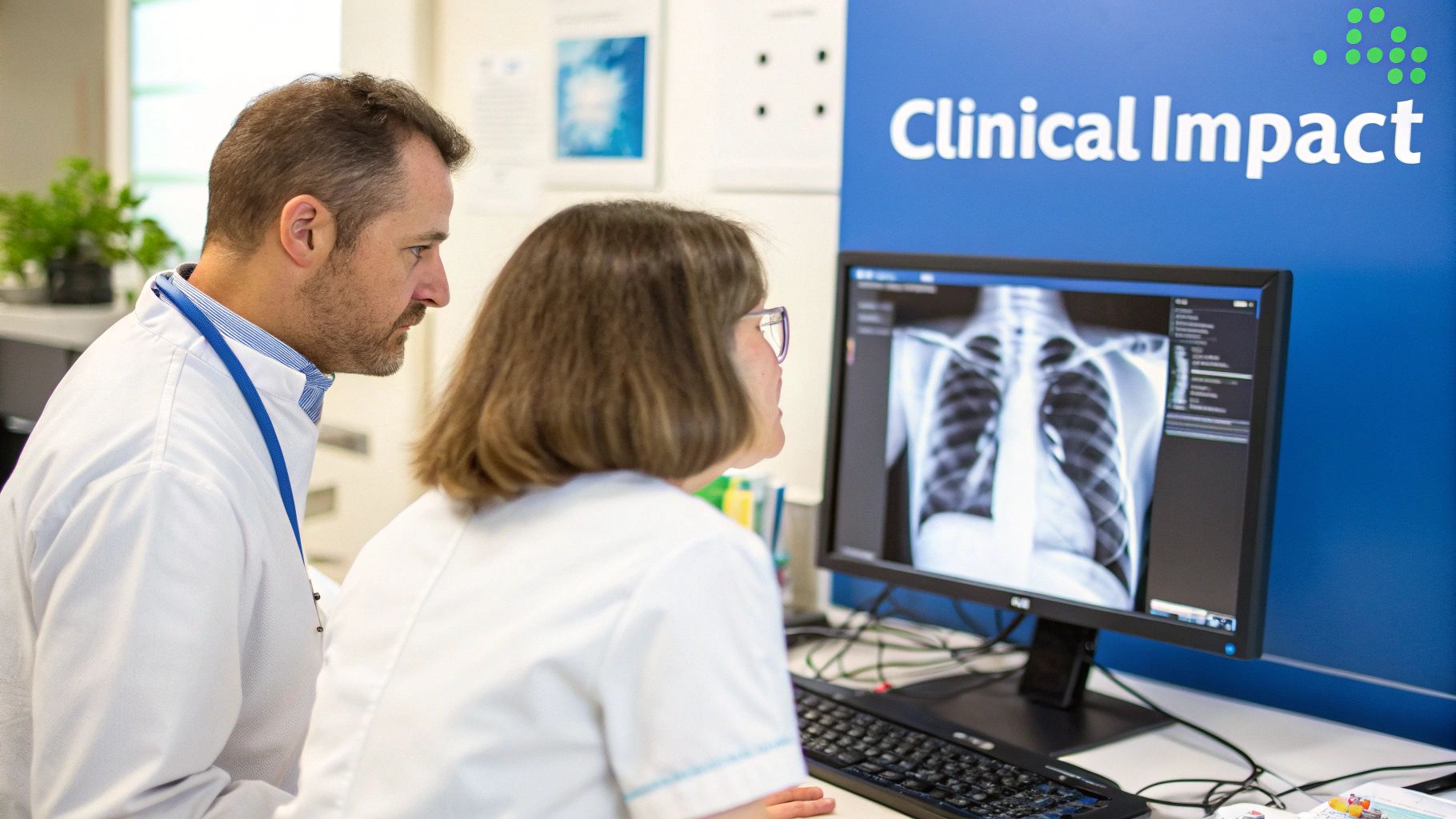
Enhancing Diagnostic Radiology Workflows
Radiology is ground zero for the impact of machine learning. Radiologists are facing a tidal wave of scans, and AI is proving to be an indispensable tool for staying afloat without sacrificing accuracy.
One of the most immediate benefits is triage. A well-trained model can analyze a head CT, instantly flag a potential brain bleed, and bump that case to the top of the worklist. This simple act of prioritization ensures life-threatening conditions get a specialist's eyes on them in minutes, not hours.
This automation also extends to detection and measurement. For example, some of the most effective AI applications for lung nodule detection can meticulously comb through thousands of CT slices to find tiny nodules a person might overlook during a long shift. It's about reducing errors and catching cancer earlier.
Given that CT imaging accounts for 37% to 40.6% of the global market, it's no surprise that this is where ML is making its biggest mark. In peer-reviewed trials, algorithms are identifying lesions with over 95% sensitivity, helping reduce radiologist workloads by a staggering 20-30%. This is especially critical in high-volume fields like oncology, a sector where AI adoption is growing at a 30.2% compound annual growth rate.
Powering Precision in Pathology
Digital pathology has swung the doors wide open for machine learning to bring a new level of precision to cancer diagnosis. For decades, pathologists have spent countless hours hunched over a microscope, meticulously examining tissue slides for cancerous cells.
Now, AI models can analyze high-resolution digital scans of those same slides to:
- Identify and count mitotic figures: A key marker for how aggressively a tumor is growing. AI does it faster and more consistently than a human can.
- Grade tumors: Algorithms assess cellular features to help classify a cancer's severity, which is fundamental to building the right treatment plan.
- Pinpoint metastases: Machine learning can spot tiny clusters of cancer cells in lymph nodes that are incredibly easy for the human eye to miss.
This isn't about replacing pathologists. It's about freeing them up to focus on the most complex diagnostic puzzles where their experience is truly irreplaceable.
One of our featured clinical speakers, a practicing oncologist, offers a concise take: "These tools change our workflow for the better. The AI provides a first pass, flagging areas of concern with incredible accuracy. This lets my team and I concentrate on interpreting complex cases and discussing treatment options with our patients."
Advancing Cardiac Imaging Analysis
In cardiology, assessing heart function means analyzing motion from sources like echocardiograms and cardiac MRIs. Historically, this has involved tedious, time-consuming manual measurements.
Machine learning is changing the game by automating these tasks with remarkable precision. Algorithms can now instantly calculate key metrics that tell the story of a patient's heart health, including:
- Ejection fraction: The percentage of blood leaving the heart with each beat.
- Strain analysis: A detailed look at how well the heart muscle is functioning.
- Wall motion abnormalities: Identifying parts of the heart muscle that aren't contracting correctly.
This automation doesn't just save cardiologists time; it delivers more consistent and reproducible measurements. That consistency is vital for tracking a patient's condition over months or years, giving clinicians a clearer picture of whether a treatment is working and empowering more informed decisions.
Navigating the Hurdles of AI Adoption
While the potential of medical imaging machine learning is enormous, the journey from a research lab to a patient's bedside is packed with serious ethical and practical roadblocks. Adopting this technology the right way means tackling these hurdles head-on to make sure innovation actually serves patients safely and fairly.
One of the biggest conversations revolves around the "black box" problem. Many of the most powerful AI models, especially deep learning networks, reach their conclusions in ways that are nearly impossible for a human to follow. This lack of transparency can really undermine a clinician's trust—doctors are, quite rightly, hesitant to act on a recommendation they can't explain or double-check.
This is where the push for explainable AI (XAI) comes in. These are systems built specifically to clarify why an algorithm landed on a particular prediction, turning complex math into insights a doctor can actually use. You can get a much deeper look into these methods in our complete guide on what explainable AI is and why it matters. Without that layer of trust, widespread adoption just won't happen.
Protecting Patients and Preventing Bias
Beyond just transparency, data privacy is a massive concern. Medical imaging datasets contain incredibly sensitive personal health information, which is protected by strict rules like HIPAA in the United States. Before any ethical AI development can even begin, it's non-negotiable that these huge datasets are completely anonymized and locked down against any potential breaches.
Then there's the equally profound challenge of algorithmic bias. At the end of the day, an AI model is only as good as the data it was trained on. If that training data mostly comes from one demographic, the final algorithm could be far less accurate for underrepresented groups, which risks either creating or worsening existing health disparities.
A featured speaker on our roster, an expert in AI ethics, frequently emphasizes this point: "Algorithmic bias isn't just a technical problem; it's a critical patient safety issue. If our AI tools don't work equally well for everyone, we risk building a future of medicine that is less equitable than the one we have today. Diligent, inclusive data curation is the only path forward."
What this means in practice is that creating fair and effective medical AI requires a deliberate, focused effort to build diverse datasets that truly reflect the full spectrum of the patient population.
The Rigorous Path to Clinical Use
Finally, even a perfectly transparent and unbiased model has to face a tough regulatory journey. In the U.S., any AI tool that will be used for diagnosis has to go through intensive validation to get clearance or approval from the Food and Drug Administration (FDA). This whole process is designed to prove the technology is both safe and effective for its specific clinical use.
That path involves a ton of testing, clinical trials, and meticulous documentation, all of which adds up to a significant investment of time and resources.
To make sense of it all, it helps to break down the primary challenges that organizations face on the road to implementation.
Key Challenges in Medical AI Implementation
Here's a breakdown of the primary ethical and regulatory hurdles facing the adoption of machine learning in medical imaging, outlining each issue and potential solutions.
| Challenge Area | Description of the Issue | Potential Mitigation Strategy |
|---|---|---|
| The 'Black Box' Problem | AI decision-making processes can be opaque, making it difficult for clinicians to trust or verify outputs. | Implement explainable AI (XAI) techniques that provide clear justifications for the model's predictions. |
| Algorithmic Bias | Models trained on non-diverse data can perform poorly on underrepresented populations, creating health inequities. | Actively curate and validate training datasets to ensure they are large, diverse, and representative of all patient groups. |
| Data Privacy & Security | Patient data used for training is highly sensitive and protected by regulations like HIPAA. | Employ robust anonymization protocols and state-of-the-art cybersecurity measures to protect all patient information. |
| Regulatory Approval | AI diagnostic tools must pass stringent validation processes with bodies like the FDA before clinical use. | Engage with regulatory experts early and design validation studies that meet or exceed established standards for safety and efficacy. |
Successfully clearing these hurdles is absolutely essential if we want to realize the full, responsible potential of machine learning in medicine. It demands a coordinated effort between developers, clinicians, ethicists, and regulators, all working toward the same goal.
Bringing AI from the Lab into the Clinic
A powerful algorithm is just a research project until it proves its worth in a real hospital. Getting a medical imaging machine learning model out of the lab is a massive undertaking, moving it beyond clean code and into the messy, complicated world of clinical workflows, patient safety, and existing hospital IT.
It’s where theoretical accuracy meets the unpredictable reality of patient care.

This transition is the ultimate test. A model that looks perfect on a curated dataset can easily stumble when it meets the sheer diversity of data from a real patient population. Success demands a careful, step-by-step approach.
From High-Quality Data to Seamless Integration
The journey begins long before a single line of code is written for the hospital's servers. It starts with the foundation of any trustworthy AI: high-quality training data. This means curating large, diverse, and meticulously anonymized datasets that truly represent the patients the tool will eventually see.
Once a model is trained and validated, the next huge hurdle is technical integration. An AI tool is completely useless if it doesn’t talk to the systems clinicians already use every single day.
This means ensuring seamless connectivity with:
- Picture Archiving and Communication Systems (PACS): This is the digital backbone of any radiology department, where all medical images live.
- Electronic Medical Records (EMRs): These systems hold the entire patient story—lab results, clinical notes, and medical history—providing critical context for any diagnostic tool.
True integration means the AI’s insights pop up directly inside the clinician’s existing workflow, not in some separate, clunky application they have to switch to.
The Gold Standard Human-in-the-Loop Systems
One of the most critical principles for safe adoption is the human-in-the-loop model. In this setup, the AI never makes the final diagnosis. Instead, it acts as an incredibly skilled assistant.
The model might flag potential abnormalities, perform rapid measurements, or prioritize urgent cases, but a human expert always, always has the final say.
This collaborative framework gives you the best of both worlds: the AI’s speed and knack for spotting subtle patterns combined with the clinician's deep medical expertise, intuition, and understanding of the individual patient. It's why these platforms are best understood as powerful clinical decision support systems that augment—not replace—human judgment.
As one of our speakers with deep implementation experience often says, "The goal is not to build an AI that replaces a doctor. The goal is to build an AI that makes a good doctor even better, more efficient, and more accurate."
This approach is absolutely vital for building trust and ensuring that patient care stays firmly in the hands of qualified professionals.
Continuous Monitoring for Lasting Accuracy
Deployment isn't a one-and-done event. It's the start of a continuous process of monitoring and refinement. A model’s performance can drift over time as imaging equipment gets updated, patient demographics shift, or new disease variants emerge.
Continuous performance monitoring is non-negotiable. It involves constantly tracking the model’s accuracy against real-world outcomes to ensure it remains reliable and safe. If performance slips, the model might need to be retrained or recalibrated with fresh data.
This global push for deployment is seeing massive investment. In fact, the Asia-Pacific region is the fastest-growing market for AI in medical imaging, with a projected growth rate of 30.8% from 2025 to 2034. This surge is driven by huge R&D investments in countries like India and China, creating a boom in tools designed to improve healthcare access.
Hearing from a speaker who has navigated this entire process—from data curation to post-deployment monitoring—provides an invaluable roadmap for any organization looking to bring these powerful tools into their own clinical environment.
Got Questions About AI in Medical Imaging? We've Got Answers.
As AI tools become more common in clinics and hospitals, it’s only natural for questions to pop up. How does this technology actually work? What does it mean for clinicians? And where is all this heading?
Getting clear, straightforward answers is the first step toward building the trust needed to bring these tools into the mainstream.
Below, we’ll tackle some of the most common questions we hear. We've also woven in the kinds of insights you’d get from the world-class experts on our speaker roster, who spend their time demystifying these topics for audiences around the globe.
What's the Difference Between AI, Machine Learning, and Deep Learning?
It helps to think of these terms like a set of nesting dolls.
- Artificial Intelligence (AI) is the biggest doll—the broad idea of making machines that can think, reason, and solve problems like humans.
- Machine Learning (ML) is the next doll inside. It’s a specific branch of AI where systems learn from data to make predictions, rather than being explicitly programmed for a task.
- Deep Learning (DL) is the smallest doll at the very center. This is a supercharged type of machine learning that uses complex, layered neural networks—inspired by the human brain—to analyze massive datasets.
When you hear about breakthroughs in medical imaging today, they are almost always powered by deep learning. Its multi-layered structure is incredibly good at spotting subtle, complex patterns in visual data like MRIs and CT scans, allowing it to "see" things that other methods might miss.
Will AI Replace Radiologists?
This is easily the most common question, and the answer you'll hear from every expert is a clear and confident "no." The goal of AI in medical imaging is augmentation, not replacement.
Think of AI as a force multiplier for human experts, not a substitute. It’s a tool designed to amplify their skills and free them up to do what they do best.
An AI can sift through thousands of chest X-rays in minutes, flagging the ten most concerning scans that need immediate attention. This lets a human radiologist focus their expertise right where it’s needed most, instead of manually working through a massive queue.
As one of our top clinical speakers often puts it: "AI is brilliant at high-volume pattern recognition. But it can't consult with a surgeon, comfort a worried patient, or apply the years of intuition needed to crack a truly bizarre diagnostic puzzle. It handles the 'what,' so we can focus on the 'why' and 'what's next.'"
This human-in-the-loop model helps reduce burnout, cuts down on tedious work, and elevates the clinician’s role to focus on critical thinking, patient care, and collaboration.
How Is a Medical AI Model Validated for Safety?
Getting an AI tool into a real clinical setting isn't easy, and for good reason. It's a massive responsibility, governed by a rigorous validation process to prove an algorithm is both safe and effective. It's not enough to be clever—it has to be reliable.
The journey to the bedside typically follows a few critical steps:
- Retrospective Validation: First, the model is tested against huge, historical datasets it has never seen before. This is where its raw performance—accuracy, sensitivity, and specificity—is measured in a controlled setting.
- Prospective Studies: Next, the model goes into a real-time clinical trial. This is the true test, measuring how it performs in an actual hospital workflow with all the messiness and unpredictability of the real world.
- Regulatory Review: Finally, the model, along with all its performance data, is submitted to a regulatory body like the Food and Drug Administration (FDA) in the US. Getting FDA clearance is the final stamp of approval, confirming the tool meets the highest standards for patient safety.
Only the most robust and trustworthy models ever make it through this gauntlet.
What Are the Biggest Barriers to Adopting AI in Hospitals?
Despite all the potential, actually getting AI integrated into a hospital is a heavy lift with some major hurdles. An expert speaker specializing in clinical implementation would point to three big challenges that pop up again and again.
First is the technical integration. A new AI tool has to play nicely with a hospital's existing IT systems, especially the Picture Archiving and Communication Systems (PACS) and Electronic Medical Records (EMR). If the software isn't seamlessly woven into the clinician’s day-to-day workflow, it simply won't get used.
Second, there are the data challenges. To build an unbiased, high-performing model, you need access to enormous amounts of diverse, high-quality, and properly anonymized patient data. Just sourcing and preparing that data is a monumental task in itself.
Finally, you have the regulatory and reimbursement hurdles. Navigating the long and complex FDA approval process is a major undertaking. On top of that, figuring out payment models and getting insurance companies to cover AI-assisted diagnostics is an ongoing challenge that’s crucial for making these tools financially viable for providers.
Ready to bring clarity and expert insight on medical imaging machine learning to your next event? At Speak About AI, we connect you with the world's leading minds in the field. Explore our roster of speakers and find the perfect expert to inspire your audience at https://speakabout.ai.